Physics of quantitative imaging systems
Development of imaging devices with enhanced quantitative capabilities through incorporation of new sensors, data acquisition protocols, imaging trajectories, and contrast mechanisms.
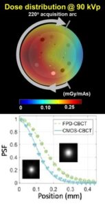
- Recent research includes optimization of ultra-high resolution CT for imaging of bone and lung microstructure, evaluation of new x-ray detector designs (including novel pixelated scintillators for high-resolution imaging), investigation of robotic x-ray devices for anatomical measurements of weight-bearing skeleton, and development of spectral imaging hardware for better quantification of bone composition.
Recent publications:
- Cao, A. Sisniega, M. Brehler, JW. Stayman J. Yorkston, J.H.Siewerdsen, W. Zbijewski, Modeling and Evaluation of a High-Resolution CMOS Detector for Cone-Beam CT of the Extremities, Medical Physics 45, 114–130, 2018
- Zhao, M. Herbst, S. Vogt, L. Ritschl, S. Kappler, JH. Siewerdsen, W. Zbijewski, Cone-Beam Imaging with Tilted Rotation Axis: Method and Application Using a Robotic Imaging System, Medical Physics, 47, 3305-3320, 2020
Image formation algorithms for improved quantitative imaging
Algorithms to process raw sensor data to generate multi-dimensional images suitable for quantitative analysis.
- Our goal is to develop
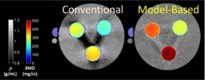 algorithms that enable robust and accurate quantification through improved handling of non-idealities in the input data, frequently by incorporating advanced system models in the image formation process. Projects in this area include reduction of blur due to patient motion to improve quantification of trabecular microarchitecture, mitigation of inaccuracies due to metal surgical hardware in Dual Energy material decomposition, fast and accurate scatter correction in CT, and model-based CT reconstruction for quantitative applications.
Recent publications:
- Sisniega, G. Thawait, D. Shakoor, J.H. Siewerdsen, S. Demehri, W. Zbijewski, Motion Compensation in Extremity Cone-Beam Computed Tomography, Skeletal Radiology, 48(12), 1999–2007, 2019
- S.Z. Liu, Q. Cao, J.H. Siewerdsen, M. Tivnan, S. Tilley II, J.H. Siewerdsen, J.W. Stayman, W. Zbijewski, Model-based dual-energy tomographic image reconstruction of objects containing known metal components, Physics in Medicine and Biology, Oct 2020.
Quantitative image analytics
 Automated approaches to extract new types of quantitative information from medical images, including validation in clinical applications.
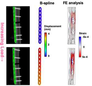
- We investigate advanced image analysis and data scientific methods to convert multi-dimensional imaging data into quantitative biomarkers for diagnosis and treatment planning. Among recent developments are automated anatomical measurements of weight-bearing joint morphology in orthopedic cone-beam CT, image-based modeling of load-induced surgical hardware deformation to assess fracture healing, and morphological analysis of 3D tumor morphology revealed by clearing microscopy.
Recent publications:
- M Brehler, Q. Cao, K.F. Moseley, G.M. Osgood, C.D. Morris, S. Demehri, J. Yorkston, J.H. Siewerdsen, W. Zbijewski, Robust quantitative assessment of trabecular microarchitecture in extremity cone-beam CT using optimized segmentation algorithms, SPIE Medical Imaging, 105781J, 2018
- Brehler, G. Thawait, Q. Cao, J. Ramsay, S. Demehri, J.H. Siewerdsen, W. Zbijewski, Atlas-based algorithm for automatic anatomical measurements in the knee, Journal of Medical Imaging, 6(2), 2019